Underwriting and Claims
  • Research and White Papers
  • July 2024
  • 7 minutes

Borderline Ovarian Tumor: Implications for critical illness and terminal illness claims

By
  • Dr. Radhika (Radi) Counsell
Skip to Authors and Experts
Doctor explaining results to woman
In Brief

Borderline ovarian tumors (BOT) can cause confusion for claims assessors. In this article from RGA's ReFlections newsletter, RGA's Dr. Radhika Counsell explains BOT diagnosis for critical illness and terminal illness claims.

Abstract

  • Borderline ovarian tumor (BOT) is an intermediate condition in the tumor spectrum.
  • It can cause confusion for claims assessors examining critical illness insurance claims, due to the cells being potentially precancerous, as well as the possibility of microinvasion and of non-invasive peritoneal implants.
  • Assessment of BOT-related terminal illness claims on life insurance policies can also be complicated, as some BOTs can transform into invasive low-grade serous carcinoma (LGSC).
  • This article explores the implications of a BOT diagnosis for critical illness and terminal illness claims, using case study examples. 


Background

Borderline ovarian tumor (BOT) comprises about 15% of all ovarian tumors.1 Clinically, these lesions are considered intermediate, between benign cystadenomas and invasive cancers, but they are not cancer. Their histological appearance is neither benign nor frankly malignant,2 and their growth rate is moderate and more controlled than ovarian cancer.

At diagnosis, BOT is usually confined to the ovary, although non-invasive peritoneal implants can develop in about one-third of cases. Cellular proliferation with BOT is greater than with benign tumors, and the appearance of the cells is more abnormal. Unlike with malignant tumors, there is no destructive stromal invasion; however, microinvasion (i.e., the presence of BOT cells beyond the outer layer of the ovary) can occur. 

According to the latest World Health Organization (WHO) Classification 2014,3 the term “borderline ovarian tumor” is interchangeable with the term “atypical proliferative tumor.” Notably, use of the phrase “tumors of low malignant potential” to describe these neoplasms is no longer recommended. 

Classification and pathology

There are six subtypes of BOT.4 The majority, about 65% to 70%, are classified as serous (meaning it arose from the serous membrane). The less common subtypes are mucinous, endometrioid, clear cell, seromucinous, and borderline Brenner type. Expert opinion is that each subtype has its own distinct biology, pathology, and molecular profile, which means no single unifying concept exists for the range of BOT subtypes.

The architecture of serous BOT is characterized by an overgrowth of cells, leading to multiple layers of stratification within the epithelial lining of the papillae and to tufting (i.e., cell detachment).5 Some tumors can exhibit a complex cribriform (also known as micropapillary) pattern. There is generally an absence of destructive stromal invasion. 

Serous BOT can also exhibit hierarchical branching of successively smaller papillae arising from larger, more centrally located papillae. Elongated micropapillae can arise directly from large central papillae as well. If this appearance is present in less than 10% of the whole tumor, the term “focal borderline change” is used as the descriptor, rather than BOT. 

If the cribriform or micropapillary architecture represents more than 10% of the tumor or 5 mm of a confluent area, some experts would label the tumor “micropapillary serous carcinoma.” Others prefer the term “serous borderline tumor with cribriform and/or micropapillary architecture,” as there is no destructive stromal invasion. In comparison with conventional serous BOT, micropapillary/cribriform serous BOTs are also more often bilateral, exophytic, at FIGO stage >1, and show invasive peritoneal implants.7 

Although BOTs do not show destructive stromal invasion, microinvasion can be seen in 10% to 20% of these tumors. Microinvasion is defined as single cells, cell clusters, or haphazard nests of cells measuring less than 5 mm that invade the stromal core of the papillae or cyst wall. If the invasive component measures more than 5 mm, the tumor should be classified as an invasive cancer. 

Although BOT has a distinct histological appearance, some of its aspects can challenge the pathologist in making a correct diagnosis.6 

Woman sitting by a waterfall reflecting on life
Explore ReFlections, the industry's premier medical underwriting publication, to learn more about key medical issues impacting the insurance industry.

Peritoneal implants

BOT can be confined to the ovary or can be seen in the peritoneum and/or the lymph nodes. Implants in the peritoneum or omentum are not uncommon in serous BOT and may be evident in about a third of BOT patients at surgery.5 The presence of implants is often associated with an exophytic neoplasm (tumor on the surface of the ovary), as fragments of tumor on the surface of the ovary can easily break off and settle in other sites away from the ovary, such as in the pelvis and abdomen.9

Previously, BOT implants were classified as either non-invasive or invasive, depending on whether destructive stromal invasion was identified. According to the 2014 WHO classification, however, the presence of invasive implants represents cancer – specifically, low-grade serous carcinoma (LGSC).3 The classification also defines the two types of non-invasive implants as 1) epithelial, in that it develops in the ovary’s epithelial tissue, or 2) desmoplastic, as it is characterized by nests of epithelial cells within a reactive stroma.

Molecular studies have found that borderline serous tumors and LGSCs carry similar mutations in the BRAF and KRAS oncogenes, implying the existence of a spectrum of change within LGSCs that develop from serous BOT.8 

Borderline ovarian tumor (BOT) is an intermediate condition in the tumor spectrum.

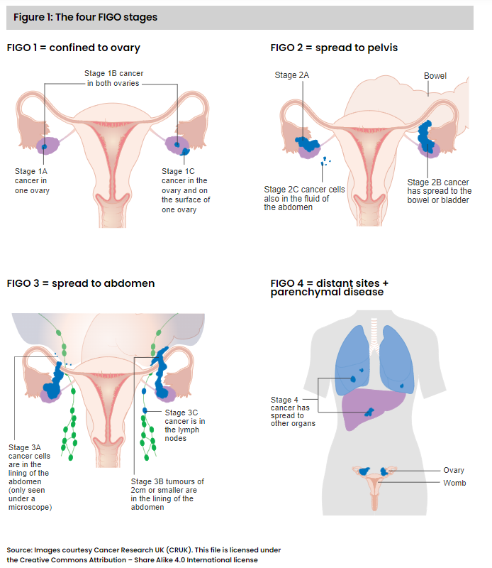
FIGO staging of ovarian tumors 

FIGO is a staging system developed by the International Federation of Gynecology and Obstetrics (FIGO) specifically for female cancers, including cervical, ovarian, uterine, endometrial, and BOT. Each FIGO stage (see Figure 1) describes the anatomical extent of tumor presence within the body. However, it does not indicate whether the tumor is an invasive cancer. 


Case Study 1: Critical illness claim 

A 40-year-old woman presented with abdominal bloating. A CT scan revealed multiple tumor masses in her pelvis and abdomen, and a blood test for presence of the protein CA125, a marker for ovarian tumors, showed a level of 85 units/ml, substantially above the normal range of 0 to 35 units/ml. The woman underwent surgery to remove the tumors, and the surgeon also biopsied suspicious nearby lymph nodes that looked larger than normal. 

The histopathology report confirmed borderline serous tumor of both ovaries and the lymph nodes. In addition, non-invasive desmoplastic implants were evident in the peritoneum of the abdomen. The FIGO stage was given as 3B, in view of the spread beyond the pelvis of tumors of up to 2 cm in diameter and the peritoneal implants. 

To qualify for a benefit, critical illness policy definitions generally require a diagnosis of malignant tumor with histological confirmation, characterized by uncontrolled growth with invasion and destruction of normal tissue. Diagnoses such as premalignant, non-invasive, carcinoma in situ (Tis) or Ta, borderline malignancy, or low malignant potential, are generally not covered. 

In this specific case, there was histological confirmation of a borderline ovarian neoplasm. Also, although there were signs of tumor within the peritoneum with non-invasive implants and presence in the lymph nodes, there was no invasive cancer. 

Non-invasive implants are not uncommon and are thought to originate from cell fragments breaking off from the BOT.9 They can generally be seen on the surface of the opposite ovary or other organs within the pelvis and abdomen as well as in the peritoneum, omentum, diaphragm, and abdominal wall. 

About one-fourth of women with BOT can also have signs of BOT cells in lymph nodes near the ovaries, but this is not due to invasive cancer and does not appear to influence long-term survival.11 

In this case, the claim was deemed to not meet the benefit definition as there was no invasive cancer. 

Clinical behavior of BOT

The majority of women presenting with BOT are of childbearing age (premenopausal) and about one-third are under age 40. Most have FIGO stage 1 disease, with tumors in both ovaries in 25% to 50% of cases.12 

The mainstay of treatment is surgery, as chemotherapy is generally not effective for BOT. As the majority of women who develop BOT want to preserve their fertility, the surgeon will usually remove only the lesion(s) and not the ovary itself. In the future, if the patient wishes, she might have a second prophylactic surgery to remove the opposite ovary and the uterus. The timing of such a surgery is usually after the woman has completed her family or after menopause.

Life expectancy for these cases is excellent.13 A meta-analysis of 97 trials with more than 4,000 women after a mean follow-up duration of about seven years found that for women with FIGO stage 1 tumors, the overall disease-specific survival rate was 99.5%. For women with tumors at more advanced FIGO stages, survival rates were influenced by the types of peritoneal implants present: 95.3% for non-invasive peritoneal implants compared with 66% for invasive implants. Neither microinvasion in the primary ovarian tumor nor the presence of BOT cells in the lymph nodes had a significant adverse effect on life expectancy, with survival rates of 100% and 98%, respectively. 

Recurrence of borderline tumors

Factors associated with borderline tumor recurrence are: FIGO stage at first diagnosis, incomplete removal of tumor(s) at surgery, and histological subtype (e.g., serous with micropapillary pattern, mucinous tumor of the peritoneum, microinvasion). Recurrent BOT can usually be treated effectively with further surgery, and the 10-year survival rate for recurrent cases is greater than 95%. 

Recurrence for BOT can manifest as another BOT, or there can be malignant transformation to LGSC. These are generally slow-growing cancers, and five-year survival rates, even for tumors of advanced FIGO stages, are around 85%.

Typically, non-serous BOTs are FIGO stage 1. Even if there is microinvasion, recurrence or metastases are uncommon. WHO 2014 argues there is little justification in calling these subtypes BOT and instead recommends calling them “atypical proliferative tumors.” (The terms “BOT” and “atypical proliferative tumor” are considered by WHO to be equivalent.) 

Critical Illness Protection: New frontiers, new customers — A look at the current global trends.

Case Study 2: Terminal illness claim

A 49-year-old woman had a first surgery for BOT four years ago. At the time, the surgeon removed both ovaries, as they were enlarged and contained tumor masses. The histopathology report noted serous BOT with micropapillae. 

She remained well until several months prior to filing her claim. She had developed bloating of the abdomen as well as intermittent vomiting and constipation. A CT scan suggested a recurrence of tumor masses at multiple sites in the abdominal peritoneum and omentum. A subsequent CT-guided biopsy confirmed recurrence of serous BOT. 

The risk factor for recurrence in her case was her subtype of BOT: serous with micropapillary pattern. 

The woman underwent surgery, and the surgeon found more tumor sites in the peritoneum than had been seen on the CT scan. In some areas, the bowel was being compressed by the tumors. Overall, it was difficult to remove all of the masses. The histopathology report showed additional areas of invasive peritoneal implants as well. 

By most policy definitions, an illness is considered terminal if the following conditions are satisfied: if there is no known cure, or if the illness being claimed for has progressed to a point where it cannot be cured and is expected to lead to death within 12 months. This means no treatments are available that may extend life expectancy, and the person has a 50% or greater chance of dying within the next 12 months. 

Many with advanced incurable cancer will continue to take medication to alleviate their symptoms. They may even have “best supportive care surgeries,” to relieve certain symptoms and improve quality of life, such as a bypass operation or stoma creation to ease the effects of a tumor blocking the bowel. (Best supportive care, whether it is medical or surgical, will not lengthen lifespan.) 

In this case, the woman had extensive invasive peritoneal implants from serous BOT and in some areas the cells had transformed to LGSC. She was experiencing bouts of pain, intestinal symptoms, and constipation from the tumors pressing the small bowel. She was prescribed analgesics, anti-emetics, and laxatives to help control her symptoms. 

Women can live with LGSC for some years, and the majority of those with advanced cases will live seven or more years. More than half with LGSC at advanced FIGO stages will live for 10 years.10 In this woman’s case, however, her surgeons had limited success removing her peritoneal tumor and were not able to bypass the areas with bowel impingement. 

Because of these factors, the woman had been advised by her doctors and surgeons that no further treatments could cure or stop her cancer progression. Chemotherapy is ineffective for BOT, and at that point, surgeons had done what was possible for her case. She therefore submitted a terminal illness claim on her life insurance policy.

This claim was deemed to meet the policy’s terminal illness criteria: She had invasive cancer for which no effective treatments were available, and her life expectancy at time of claim was 12 months or less. 

Conclusions

BOTs are neither benign tumors nor invasive cancers, although microinvasion of the stroma can be seen in some cases. 

The first case study highlights that BOTs can cause non-invasive implants within the peritoneum and that areas of BOT can develop in the lymph nodes. Although the extent of BOT warranted assignment of FIGO stage 3B, that does not indicate the tumor was an invasive malignancy. In this case, it was a BOT, and therefore did not meet the definition for an invasive cancer. 

The second case study highlights that recurrence can be due to transition of a BOT condition to LGSC. This is an invasive cancer which usually grows slowly. Even for advanced FIGO stage neoplasms or multiple recurrences, life expectancy for those with LGSC can be as much as seven to 10 years. Surgery is the mainstay of treatment for LGSC, as chemotherapy does not work very well. In this specific case, there were also signs of bowel obstruction due to compression from tumor masses that could not be removed. It was expected that she would have a few months of life left, so this was a valid terminal illness claim. 

BOT can impose challenges for the claims assessor when adjudicating applications for critical illness and terminal illness. Evaluating the histopathology report and clinical details will help the assessor make the most appropriate decision. 


More Like This...

Meet the Authors & Experts

Dr. Radhika Counsell
Author
Dr. Radhika (Radi) Counsell
Consulting Medical Officer, RGA UK

References

  1. https://doi.org/10.1002/ijc.23724
  2. https://doi.org/10.1016/j.humpath.2004.03.003
  3. https://link.springer.com/article/10.1007/s00428-016-2040-8
  4. https://link.springer.com/article/10.1007/s00428-016-2040-8
  5. https://doi.org/10.1016/S0893-3952(22)04457-X
  6. https://academic.oup.com/oncolo/article/17/12/1515/6403316#
  7. https://doi.org/10.1016/j.jogoh.2020.101965
  8. https://doi.org/10.1097%2FPAP.0b013e3181b4fffa\
  9. https://doi.org/10.1155/2023/4845887
  10. https://pubmed.ncbi.nlm.nih.gov/35204549/
  11. https://doi.org/10.1016/S1470-2045(11)70288-1
  12. https://doi.org/10.1016/j.ygyno.2005.09.021
  13. https://doi.org/10.1053/hp.2000.8048